Simple squamous epithelium slide 400x offers a captivating glimpse into the microscopic world, revealing the intricate structure and functions of this specialized tissue. Its layers, staining techniques, and clinical applications provide valuable insights into the health and well-being of living organisms.
Composed of thin, flattened cells, simple squamous epithelium forms a delicate barrier lining various body cavities and blood vessels. This introductory paragraph delves into the fascinating characteristics of this tissue, setting the stage for an in-depth exploration of its structure, staining methods, and clinical significance.
Structure of Simple Squamous Epithelium Slide 400x

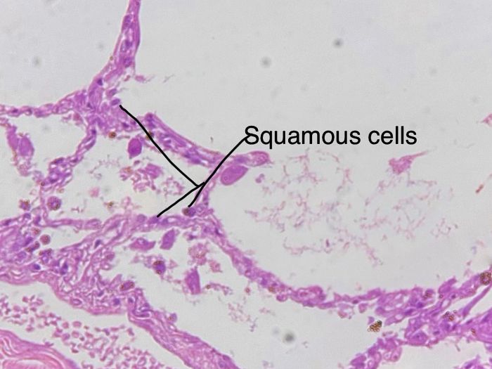
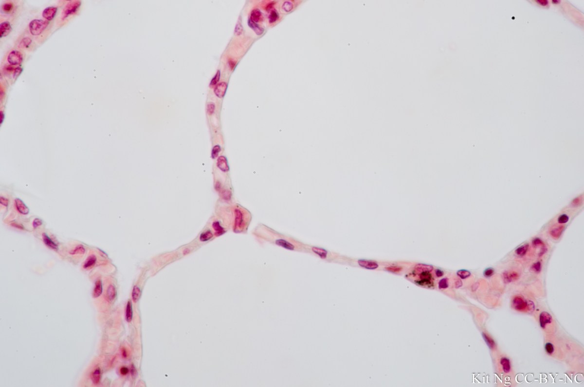
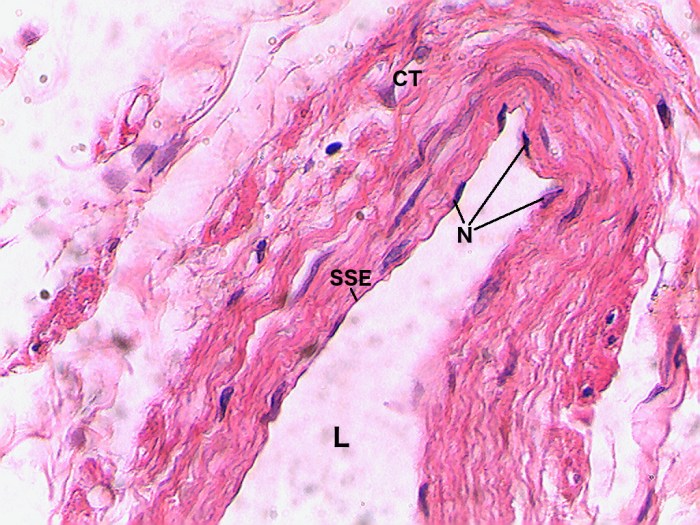
Simple squamous epithelium is a thin, single-layered epithelium that lines the surfaces of blood vessels, alveoli, and other organs. It is composed of flat, polygonal cells that are closely packed together. The cells are joined by tight junctions, which prevent the passage of molecules between them.The
structure of simple squamous epithelium is well-suited for its function. The thin, single-layered structure allows for the rapid diffusion of gases and other molecules across the epithelium. The tight junctions prevent the leakage of molecules between the cells, which is essential for maintaining the integrity of the blood vessels and alveoli.

Staining Techniques for Simple Squamous Epithelium Slide 400x

There are a variety of staining techniques that can be used to visualize simple squamous epithelium. The most common technique is hematoxylin and eosin (H&E) staining. H&E staining stains the nuclei of the cells blue and the cytoplasm pink. This allows for the visualization of the cell nuclei and the cytoplasm, as well as the tight junctions between the cells.Another
staining technique that can be used to visualize simple squamous epithelium is periodic acid-Schiff (PAS) staining. PAS staining stains the carbohydrates in the cells red. This allows for the visualization of the glycocalyx, which is a layer of carbohydrates that coats the surface of the cells.

Clinical Significance of Simple Squamous Epithelium Slide 400x

Simple squamous epithelium is used to diagnose and treat a variety of diseases. For example, simple squamous epithelium is used to diagnose lung cancer, mesothelioma, and other diseases of the lungs. It is also used to diagnose kidney disease, liver disease, and other diseases of the kidneys and liver.Simple
squamous epithelium is also used to treat a variety of diseases. For example, simple squamous epithelium is used to treat burns, wounds, and other injuries. It is also used to treat diseases of the lungs, kidneys, and liver.
| Disease | Symptoms | Treatment |
|---|---|---|
| Lung cancer | Cough, shortness of breath, chest pain | Surgery, chemotherapy, radiation therapy |
| Mesothelioma | Chest pain, shortness of breath, fatigue | Surgery, chemotherapy, radiation therapy |
| Kidney disease | Swelling, fatigue, high blood pressure | Dialysis, kidney transplant |
| Liver disease | Jaundice, fatigue, abdominal pain | Liver transplant, medication |
Preparation of Simple Squamous Epithelium Slide 400x

The preparation of a simple squamous epithelium slide 400x requires the following materials:* Simple squamous epithelium tissue
- Glass slide
- Coverslip
- Hematoxylin and eosin (H&E) stain
- Microscope
The procedure for preparing a simple squamous epithelium slide 400x is as follows:
- Place a drop of simple squamous epithelium tissue on a glass slide.
- Spread the tissue out evenly over the slide.
- Allow the tissue to dry.
- Add a drop of H&E stain to the tissue.
- Allow the stain to sit for 5 minutes.
- Rinse the slide with water.
- Dry the slide.
- Place a coverslip over the tissue.
- Examine the slide under a microscope at 400x magnification.
FAQ
What is the function of simple squamous epithelium?
Simple squamous epithelium acts as a thin, permeable barrier that facilitates the exchange of substances between different body compartments.
How is simple squamous epithelium stained for microscopy?
Common staining techniques include hematoxylin and eosin (H&E), which highlight the nuclei and cytoplasm, respectively.
What are the clinical applications of simple squamous epithelium slides?
These slides aid in the diagnosis of various diseases, including respiratory infections, urinary tract infections, and certain types of cancer.